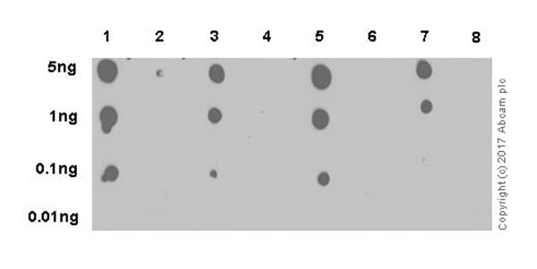


Try several different lengths of exposure. Incubate with ECL reagent for 1 min, cover with Saran wrap (remove excessive solution from the surface), and expose X-ray film in the dark room.Wash three times with TBS-T (1 x 15 min and 2 x 5 min), then once with TBS (5 min).Ensure you use an optimized protocol to give the antibody the best chance of passing the validation process. See our sample preparation guides for western blot or ELISA. For optimum antibody dilution, follow the manufacturer's recommendation. For example, an antibody that recognizes the protein only in its native form should not be used on samples using denaturing conditions, such as western blot. Incubate with secondary antibody conjugated with HRP for 30 min at room temperature.Wash three times with TBS-T (3 x 5 min).Incubate with primary antibody (0.1–10 µg/mL for purified antibody, 1:1,000–1:100,000 for antisera, 1:100–1:10,000 for hybridoma supernatant) in BSA/TBS-T for 30 min at room temperature.Block non-specific sites by soaking in 5% BSA in TBS-T in a 10 cm Petri dish (30 min to 1 h at room temperature).Minimize the area that the solution penetrates (usually 2–4 mm diameter) by applying it slowly. Using a narrow-mouth pipette tip, spot 2 µL of sample onto the nitrocellulose membrane at the center of the grid.Proteins come up as clear zones in a translucent blue background. Wash the gels briefly in de-ionized water, and view them against a dark-field background. Draw a grid by pencil to indicate the region you are going to blot. Briefly rinse freshly-electrophoresed gels in distilled water (30 sec maximum) and then transfer to a solution of 0.3 M CuCl 2 for 515 min. Have the nitrocellulose membrane ready.

For a 1x solution, mix 1 part of the 10x solution with 9 parts distilled water and adjust pH to 7.6 again. Nitrocellulose membrane (BIO-RAD, Trans-Blot, etc.) Add distilled water to a final volume of 1 L. A technique for detecting, analyzing and identifying proteins, similar to the western blot technique but differing in that protein samples are not separated electrophoretically but are spotted through circular templates directly onto the membrane or paper substrate.Ĭoncentration of proteins in crude preparations (such as culture supernatant) can be estimated semiquantitatively by using the dot blot method if you have both purified protein and specific antibody against it.


 0 kommentar(er)
